Insect Head Anatomy
The insect head is the foremost part of the body, composed of 6 fused segments, which collectively form a head capsule. The head is responsible for sensory perception, feeding, and house the brain. The head is connected to the thorax via a flexible region called the neck or cervix.
Head Segments:
- Procephalon: This is the region that includes sensory organs, compound eyes, and simple eyes.
- Gnathocephalon (Mouth Region): Includes mouthparts like mandibles and maxillae that are responsible for feeding.
Head Capsule:
- The head capsule is sclerotized, which means it is hardened by the presence of sclerites (cuticular plates or areas) fused together. The portion of the head capsule excluding the appendages is called the cranium.
- Inside the head is an endoskeletal structure known as the tentorium, which supports the brain and serves as a rigid attachment point for muscles that control the mandibles and mouthparts.
Head Appendages and Segments:
- Pre-Antennary Segment (Segment I): Contains compound eyes and three simple eyes (ocelli).
- Antennary Segment (Segment II): Holds the antennae.
- Mandibular Segment (Segment IV): Contains the mandibles used for biting and chewing.
- Maxillary Segment (Segment V): Contains the maxillae (paired appendages involved in feeding).
- Labial Segment (Segment VI): Contains the labium (lower lip of the insect).
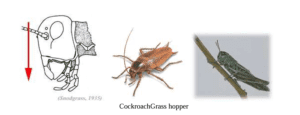
Types of Head Position:
The head’s orientation relative to the body can vary, affecting the projection of the mouthparts. The classification of head position depends on the alignment and projection of the mouthparts:
- Hypognathous: The head is vertical and positioned at a right angle to the body. The mouthparts are directed downward. Common in Orthopteroid insects like grasshoppers and cockroaches.
- Prognathous: The head remains aligned with the body, and the mouthparts project forward. Found in Coleopteroid insects like beetles.
- Opisthognathous: Similar to prognathous, but the mouthparts point backward and are positioned between the forelegs. Common in Hemipteroid insects like bugs.
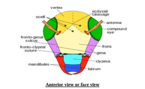
Sclerites and Sutures of the Head:
The head capsule consists of several sclerites that are fused and separated by sutures. Sutures are grooves or lines that provide mechanical support to the head and facilitate moulting.
Common Sclerites:
- Labrum: The upper lip of the mouth, attached to the clypeus.
- Clypeus: Situated above the labrum, separated by the frontal-clypeal suture.
- Frons: The unpaired area located between the arms of the epicranial suture.
- Gena: Area between the compound eyes and mandibles, separated from the frons by the frontogenal suture.
- Epicranium: The upper region of the head extending from the frons to the neck.
- Vertex: The upper portion of the epicranium, located between the compound eyes.
- Occiput: The posterior part of the head, just before the neck.
- Post occiput: The extreme posterior part, just before the neck region.
- Occular Sclerites: Cuticular rings around each compound eye.
- Antennal Sclerites: Sclerites around the base of the antennae.
Head Sutures:
Sutures are lines or ridges separating different sclerites:
- Clypeolabral Suture: Between the clypeus and labrum.
- Clypeofrontal Suture: Between the clypeus and frons.
- Epicranial Suture: A Y-shaped suture marking the boundary between the frons and epicranium. It is also known as the ecdysial line, as it splits during moulting.
- Occipital Suture: A U-shaped suture between the epicranium and occiput.
- Post Occipital Suture: Marks the boundary between the head and neck; it is the only real suture.
- Genal Suture: Located on the lateral sides of the head, at the gena.
- Occular Suture: A circular suture around the compound eyes.
- Antennal Suture: A depressed ring around the antennal socket.